EEG Imaging Method Maps Motor Learning Brain Changes
A smarter EEG brain imaging approach tracks how the cortex rewires itself as people learn new movements, sharpening scalp EEG's view of motor plasticity.
Updated
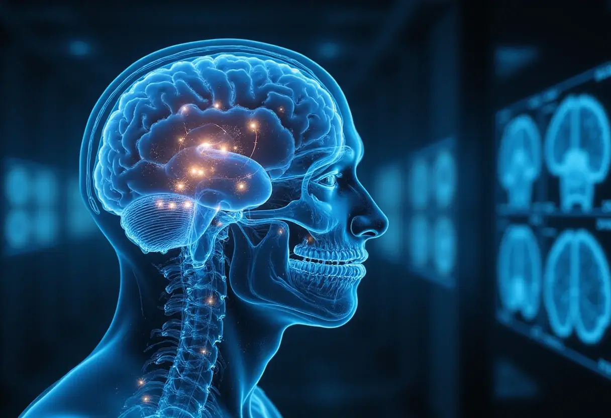
A new EEG brain imaging approach can track how the human brain rewires itself as a person learns new movements, according to a report published by Bioengineer.org. The work centers on improving the spatial and temporal resolution of scalp electroencephalography (EEG), the non-invasive technique that records electrical activity through electrodes placed on the scalp.
For clinical neurophysiology readers, the claim matters because conventional scalp EEG offers excellent millisecond-scale timing but poor localization of the cortical generators behind the signals. A method that sharpens EEG-based imaging of motor regions could give epilepsy monitoring units and motor-control laboratories a cheaper, more portable alternative to magnetoencephalography (MEG) or functional MRI when studying motor cortex plasticity.
What does the study track?
The report describes tracking neuroplasticity — the structural and functional reorganization of neural circuits — during motor learning. Motor learning tasks, such as practicing a new sequence of finger movements, are known from prior imaging work to change activity patterns in the sensorimotor cortex over time. The new approach aims to observe that rewiring directly from EEG recordings rather than inferring it indirectly.
The Bioengineer.org report does not specify the size of the participant cohort, the exact montage, or whether the method has been validated against intracranial recordings. Readers should treat the technical claims as described in the source rather than peer-reviewed specifications.
Investigational, not clinical
No statement in the report indicates FDA clearance or regulatory review for the method. The work should be read as investigational research into signal processing and source-imaging techniques for EEG, not as a diagnostic device ready for the reading room.
The report also does not distinguish between human and animal data explicitly, though the framing around learning new movements refers to human motor learning.
Why it matters for the lab
If the approach holds up, potential applications include:
- Tracking motor recovery after stroke in rehabilitation settings using bedside EEG rather than scanner time
- Providing neurofeedback (real-time display of brain activity to the participant) grounded in motor-cortex signals
- Offering a low-cost tool for studying skill acquisition in sports and clinical motor disorders
Those applications remain prospective. The source report presents the method as a demonstration of what smarter EEG imaging can achieve in following brain changes during learning.
The report suggests the technique could make studies of brain plasticity more accessible by relying on widely available EEG hardware. Whether it reaches the specificity of ECoG (electrocorticography, electrode grids placed directly on the cortical surface) or MEG will depend on forthcoming validation studies.
Based on
Source: Google News: EEG.
Priya Raman
Show full bio
Market editor covering regulation, policy and their impact on Clinical neurophysiology and EEG interpretation at EEG Signal.