Intracranial BCI Drives Speaking Avatar for Paralyzed Patient
An ECoG-based brain-computer interface decoded a paralyzed patient's attempted speech into an avatar's voice and facial gestures, in investigational human work reported by Medical Xpress.
Updated
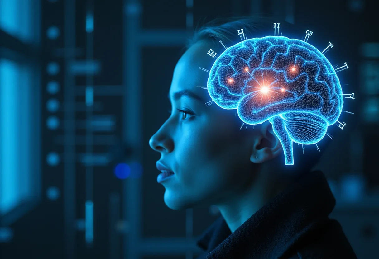
A brain-computer interface recorded directly from the cortical surface of a woman with severe paralysis and converted her attempted speech into audible output on a digital avatar — complete with facial expressions, according to coverage by Medical Xpress.
The system relies on electrocorticography (ECoG), not scalp EEG. A high-density electrode array implanted over the speech-related cortex feeds signals to a decoding pipeline that translates neural activity into text, synthesized speech, and commands for an animated avatar's mouth and facial movements. This is intracranial, investigational human research — not an FDA-cleared device, and not a commercial product.
The participant, left paralyzed and unable to speak by a brainstem stroke, achieved decoding performance that far exceeded earlier generations of speech BCI, with vocabulary handled at scale and latency low enough to sustain conversational rhythm. The avatar element is the distinguishing feature: rather than returning only text or a synthetic voice, the interface reconstructs visible orofacial gestures, aiming to restore not just communication but the social dimension of face-to-face speech.
For clinical neurophysiologists, several points deserve attention. First, the signal source. ECoG arrays over sensorimotor and language cortex carry far more spectral and spatial detail than scalp recordings, and the reported results should not be read as an expectation for extracranial montages. Scalp EEG-based speech decoding remains far more limited in bandwidth, vocabulary size, and speed. Second, the task structure. The participant attempted — not imagined — speech, engaging the same motor networks a clinical EMG or EP study would probe in an intact system, which is what makes the decoding stable enough for real-time use.
Third, the training burden. Systems of this class require extensive per-participant calibration, with models mapping electrode-level features to phonemes, words, and facial-action units. The reported setup decoded both the audio channel and the visual gesture channel, meaning two output streams derived from overlapping but partially distinct cortical populations.
The distinction between modalities matters for how readers of this site contextualize the claim. This is not mind reading, and the coverage does not claim it. The decoder outputs trained speech categories and gesture mappings from an implanted array in a single highly engaged participant after a stroke. Generalization across etiologies — ALS, traumatic injury, cerebral palsy — across electrode placements, and across longer implantation windows remains untested at scale.
For monitoring-unit and reading-room practice, the immediate relevance is indirect but real. Patients and families will increasingly arrive with expectations shaped by this class of headline. Clinicians fielding those questions should be able to state plainly what the technology is: an investigational, implant-based, participant-specific decoder that restored a form of speech output for one individual, at a level of fluency that earlier systems had not reached.
The work also signals where the field is moving: from single-output channels toward multimodal restoration, in which a BCI does not stop at text on a screen but drives voice, facial expression, and gesture together. Whether that multimodal approach survives translation beyond the research lab — with device longevity, wireless transmission, and regulatory pathways still unresolved — is the question that will determine when, or whether, such systems reach clinical use beyond a handful of study participants.
Based on
Marcus Bennett
Show full bio
Staff writer covering technology and product news in Clinical neurophysiology and EEG interpretation at EEG Signal.