Neurofeedback & Neuromodulation
MEG Neurofeedback Modulates Insula Activity in Double-Blind Trial
A double-blind randomized crossover trial in Nature shows participants can volitionally modulate insula activity using real-time neurofeedback from an MEG-based brain-machine interface.
Updated
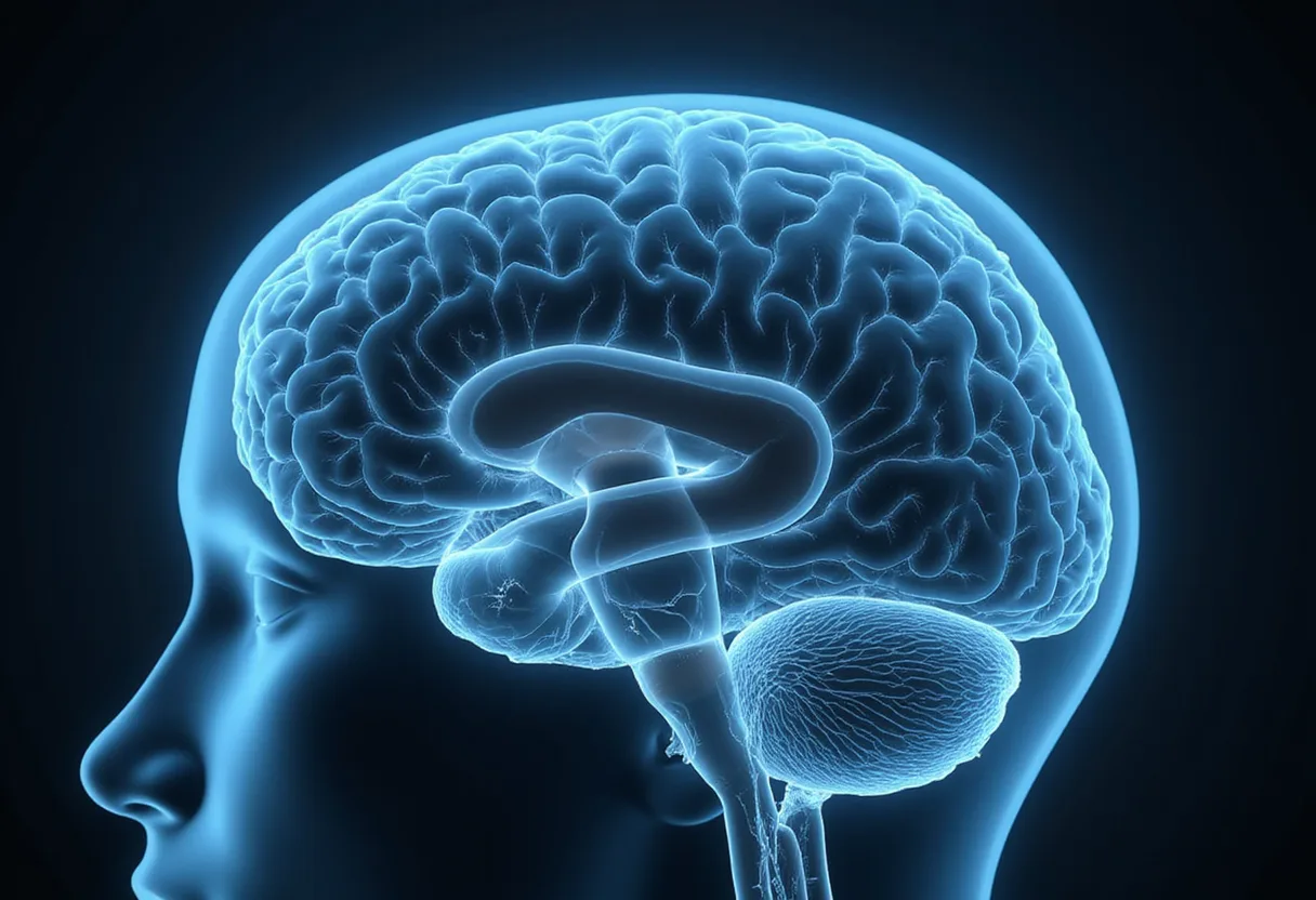
A double-blind, randomized controlled crossover trial published in Nature demonstrates that healthy participants can volitionally modulate activity in the insula using neurofeedback delivered through a magnetoencephalography-based brain-machine interface.
The study addresses a question that has constrained the neurofeedback field for years: whether regionally specific self-regulation of deep cortical structures is achievable when the signal is captured non-invasively and fed back in real time. The insula, a hub for interoception, emotional awareness, and autonomic integration, sits far enough from the cortical surface that scalp EEG struggles to resolve its activity with confidence. The investigators chose MEG for precisely this reason. MEG detects magnetic fields generated by neural currents with millisecond temporal resolution and, critically, better sensitivity to sources in the insular region than electrode-based scalp recordings.
The trial used a crossover design, meaning each participant completed both the active neurofeedback condition and a control condition, with the order randomized. This structure strengthens causal inference because each participant serves as their own control, damping inter-individual variability in baseline insula activity and in intrinsic aptitude for self-regulation tasks. The double-blind procedure means neither participants nor the experimenters delivering the sessions could distinguish active from control feedback, a safeguard against the expectancy effects and unintentional cueing that have historically inflated neurofeedback claims.
Here is how the intervention worked in principle. Participants lay in the MEG system while their insular activity was decoded in real time from the sensor-level magnetic signals. That decoded activity was then translated into a visual signal — a bar, a shape, or a comparable display — that rose or fell with the moment-to-moment activity of the target region. Participants were instructed to drive the display upward using whatever mental strategy they found effective. Over repeated training runs, the brain-machine interface closed the loop: neural activity produced feedback, and feedback guided subsequent neural activity.
The crossover results indicated that participants receiving active MEG-based feedback modulated insular activity beyond what they achieved under the control condition. Because the design was double-blind and randomized, the finding cannot be readily attributed to placebo expectancy alone — a methodological bar that much of the earlier neurofeedback literature fails to clear.
For clinical neurophysiology practice, the implications are indirect but concrete. This is investigational research, not an FDA-cleared application, and no diagnostic or therapeutic claim transfers to the reading room today. Even so, the study offers a methodological template that monitoring units and research labs can note: real-time source estimation from MEG, a target region defined anatomically rather than by convenience, a crossover design with blinding, and outcomes reported against the modulation actually achieved rather than subjective report alone.
The distinction from EEG-based neurofeedback matters for readers who work with scalp systems. MEG-based feedback rests on source localization of deep activity that scalp EEG montages cannot isolate cleanly, so the results do not automatically justify claims that standard 19-electrode or even high-density EEG protocols can train the insula specifically. Where conventional neurofeedback targets band power at scalp electrodes, this protocol targeted a defined cortical structure through an inverse-model decode. That difference in measurement fidelity is the load-bearing element of the study.
The double-blind crossover format also sets a benchmark for trial design in the neurofeedback and BCI space more broadly. Studies of neurofeedback for ADHD, epilepsy self-regulation training, and motor rehabilitation BCI have often relied on waitlist or non-blinded comparators. A crossover design with blinded sham feedback, as used here, provides a more rigorous answer to the question of whether the feedback itself — rather than the therapeutic encounter — drives the learned modulation.
The findings also bear on the debate over what neurofeedback trains. If participants can learn to regulate insular activity without electrodes on the brain, the learned signal must be accessible to non-invasive measurement and must be under at least partial volitional control. The insula's role in interoception and emotion regulation gives the result potential relevance to conditions where that region shows altered activation, including anxiety disorders and chronic pain, though the trial itself involved modulation in the study cohort, not treatment of any clinical population.
The limitations follow directly from the source. The study demonstrates modulation, not a clinical endpoint. The hardware — a whole-head MEG system — is expensive, immobile, and available only in specialized centers, which constrains any near-term clinical deployment. Sustained regulation after the training sessions, generalization to other regions or tasks, and durability of the effect all remain open questions that future trials must address.
What the trial establishes, on its own terms, is that a blinded, randomized, crossover design can demonstrate volitional modulation of insular activity through an MEG-based brain-machine interface. Whether the approach can move from the shielded MEG room into practical neurofeedback protocols will depend on the replication and durability data that investigators publish next.
Based on
Source: Google News: Neurofeedback.
Tom Whitfield
Show full bio
Senior reporter covering people, launches and day-to-day practice in Clinical neurophysiology and EEG interpretation at EEG Signal.