Scalp EEG Microstates Altered in Etomidate Use Disorder, Study Finds
A cross-sectional study reports altered scalp EEG microstates and elevated impulsivity in individuals with etomidate use disorder, an association within reach of standard clinical EEG equipment.
Updated
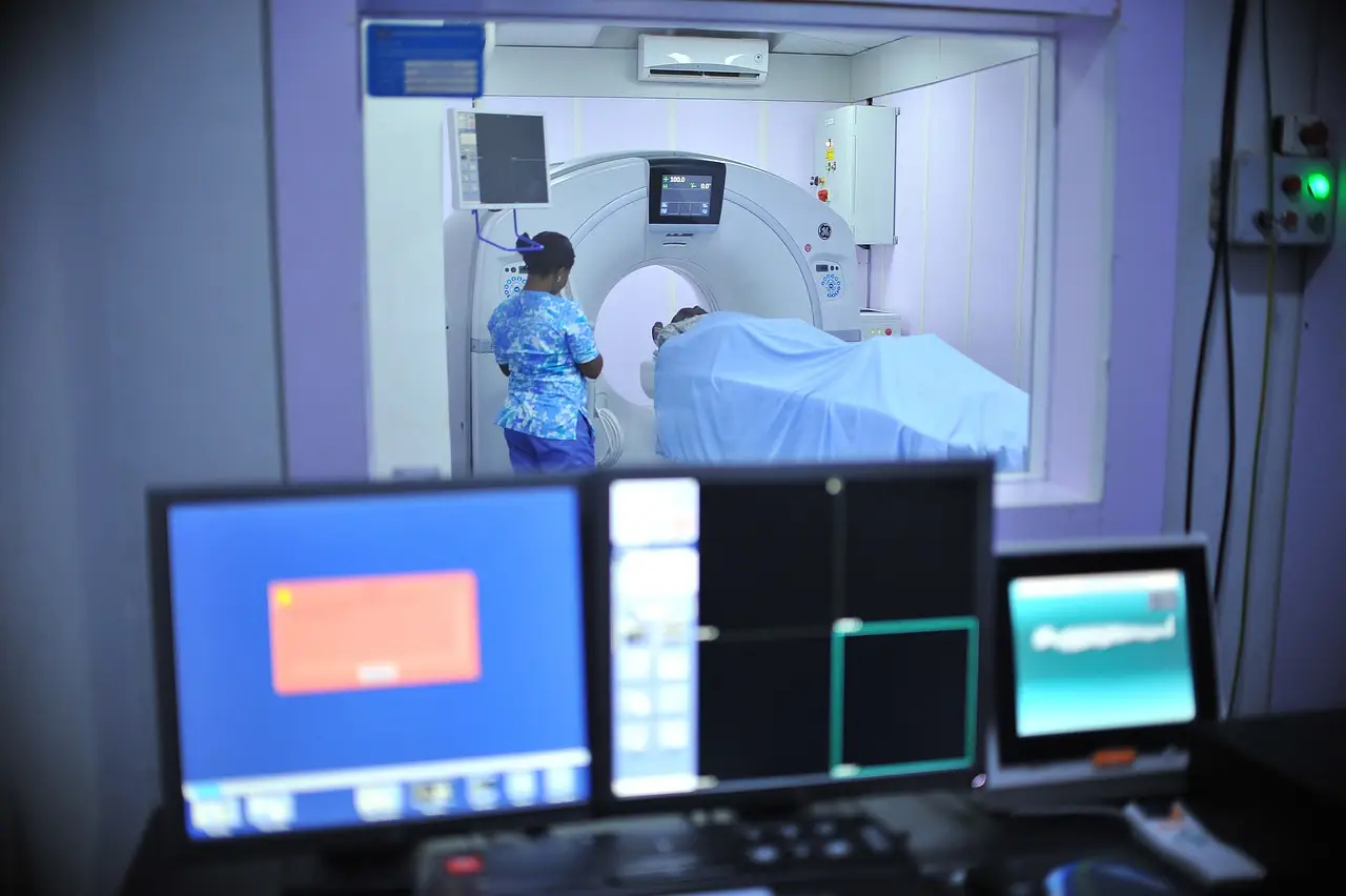
A cross-sectional study has linked etomidate use disorder with impulsivity and altered scalp EEG microstates, adding a measurable neurophysiological signature to a substance misuse problem that clinicians largely track through behavioral reports.
The study examined resting-state scalp EEG microstates — brief, stereotyped periods of quasi-stable global field topography that typically last 60–120 milliseconds and repeat across the recording. The researchers compared individuals with etomidate use disorder against controls and reported that the disorder group showed altered microstate parameters together with elevated impulsivity scores.
What are EEG microstates, and why do they matter here?
Microstate analysis condenses multichannel scalp EEG into a sequence of discrete topographic templates, classically labeled A through D. Researchers quantify how long each template persists, how often it recurs and how large its contribution is to the total signal.
These metrics index the dynamic coordination of large-scale cortical networks. Changes in microstate duration or occurrence have appeared in studies of schizophrenia, depression and other substance use disorders. The new etomidate findings place an anesthetic misuse syndrome within that same measurement framework.
The study used scalp EEG, a non-invasive modality already standard in clinical neurophysiology laboratories. That choice matters: any microstate markers confirmed in larger cohorts could be collected with existing hospital equipment rather than specialized imaging.
What did the impulsivity data show?
The authors administered validated impulsivity assessments alongside the EEG recordings. Participants with etomidate use disorder scored higher on impulsivity measures than controls, and the study design allowed the researchers to examine impulsivity and microstate metrics within the same individuals rather than inferring the relationship across separate samples.
Because the design is cross-sectional, the data cannot establish whether the EEG differences preceded the disorder, emerged from chronic etomidate exposure, or reflect a third factor. The study demonstrates an association, not a causal pathway.
How should reading rooms and monitoring units read this?
For clinical neurophysiologists, the practical takeaway is that chronic etomidate exposure now joins the list of substances with reported resting-state EEG microstate alterations. Laboratories running quantitative EEG pipelines could incorporate microstate metrics when they record patients with a history of anesthetic misuse, though no diagnostic claim is justified from a single cross-sectional study.
Etomidate itself remains an FDA-cleared anesthetic induction agent; its misuse represents an off-pattern use problem documented primarily in case reports and, now, controlled EEG studies such as this one.
The findings are limited by the cross-sectional design and by whatever cohort-size constraints the source study carries. Independent replication in larger, longitudinal samples will need to establish whether the microstate signature tracks disorder severity or recovery before it can inform monitoring protocols.
If replication holds, scalp EEG microstate analysis could offer monitoring units an objective, low-cost marker of the neurophysiological changes that accompany etomidate use disorder.
Based on
Source: Google News: EEG.
Tom Whitfield
Show full bio
Senior reporter covering people, launches and day-to-day practice in Clinical neurophysiology and EEG interpretation at EEG Signal.