Ictal SPECT Retains a Role in Epilepsy Presurgical Workup, Review Finds
A Current Medical Issues review re-examines ictal brain SPECT as an adjunct in presurgical epilepsy evaluation, focusing on MRI-negative candidates and injection-timing limits.
Updated
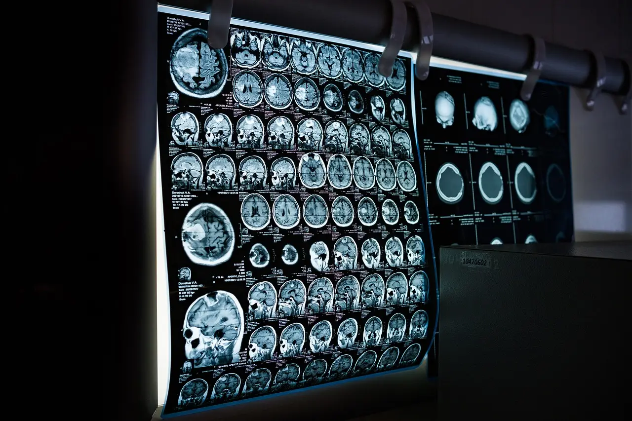
A review published in Current Medical Issues re-examines the role of ictal brain single-photon emission computed tomography (SPECT, a nuclear-medicine perfusion imaging technique) in the presurgical evaluation of patients with drug-resistant epilepsy, at a time when most epilepsy centers lean heavily on scalp video-EEG and MRI.
The paper addresses a persistent clinical gap: a substantial share of surgical candidates are MRI-negative, and non-invasive electroclinical data alone frequently fail to localize the seizure-onset zone in these patients. Ictal SPECT — a perfusion scan injected during a seizure, capturing the hyperperfused region at onset — is positioned as an adjunct that can change the trajectory of the workup, including the placement of intracranial electrodes.
What does ictal SPECT add to the EEG reading room?
For the clinical neurophysiologist, the review's framing is familiar. Scalp video-EEG with a standard montage defines the electroclinical phenotype but often produces a lateralization or a broad localization, not a resective target. Structural MRI, where concordant, narrows the hypothesis. Where those two modalities disagree or the MRI is unremarkable, ictal SPECT offers an independent, seizure-state map of cerebral perfusion that can guide — or argue against — Phase II monitoring with intracranial EEG.
The technique's well-known constraint is timing. The tracer must be injected as close to seizure onset as possible; the perfusion pattern becomes less specific as the seizure propagates and into the postictal window. This places a practical burden on the monitoring unit: injection protocols depend on staff readiness, ictal identification on the EEG review, and seizure duration long enough to permit injection before resolution.
The review situates ictal SPECT alongside other functional and structural adjuncts used in presurgical protocols, including interictal SPECT for comparison and, in many centers, subtraction techniques that co-register ictal and interictal perfusion images with the patient's MRI. These are established methods in the field, and the paper's contribution is a synthesis of when the modality earns its place rather than an endorsement of new hardware or software.
How should monitoring units read this?
The practical question the review raises for epilepsy units is what ictal SPECT changes for the MRI-negative candidate. The answer it supports is procedural: where scalp EEG and imaging leave the onset zone uncertain, a well-timed ictal injection can justify, tailor or redirect intracranial implantation — the decision point at which most presurgical evaluations succeed or stall.
The evidence base discussed is human clinical data from established presurgical practice; no new device claims, animal work or investigational technology figure in the paper. Readers should treat the review as guidance on existing, widely available nuclear-medicine methodology, not as a report on a novel FDA-cleared system.
Where the field goes from here
The article arrives as centers weigh how much of this workflow newer imaging and source-estimation methods can absorb, a question it does not settle. What it does reinforce is that, for the drug-resistant, MRI-negative population, seizure-state perfusion imaging remains part of a rational pathway from non-invasive recording to implantation — and that the deciding variable is usually the discipline of the injection protocol in the monitoring unit, not the scanner itself.
Based on
Source: Google News: Electroencephalogram.
Olivia Hart
Show full bio
Correspondent covering people, launches and day-to-day practice in Clinical neurophysiology and EEG interpretation at EEG Signal.